Taking about spinal cord MRI
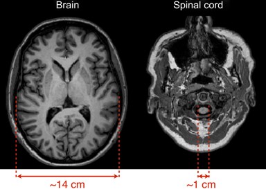
Spinal cord is the most important interface of sensory and motor signals between brain and body. We can fully understand the input and output mechanism of sensory and motor signals in central nervous system by studying spinal cord function. In addition, as the transmission pathway of somatosensory and motor signals, if the spinal cord and related nerves are injured or have diseases, the sensory and motor functions of the tissues and organs dominated by them will be abnormal. We study and understand that the role of the human spinal cord in sensory perception (such as pain and itch) and movement is often limited by the special physiological structure of the spinal cord. Spinal MRI is less common than brain imaging. The biggest reason may be that imaging on this small structure, which constantly moves with the physiological rhythm, is very challenging.
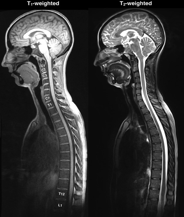
Nowadays, most of the brain imaging methods can be realized in the spinal cord, such as structural imaging (T1, T2), functional imaging (T2 *), diffusion imaging (DTI, DWI), magnetic resonance spectroscopy (MRS) and other common imaging methods used in clinical and scientific research. Spinal structural imaging is often used in the diagnosis of spinal cord lesions and the location of lesions in clinical auxiliary diagnosis. In basic scientific research, it is most commonly used in the registration of images (such as functional images and DTI). Structural images can be used to obtain quantitative indicators such as spinal cord cross-sectional area, gray matter area and length through later analysis. These indicators can be used in the research related to spinal cord plasticity changes, and can also be used In spinal cord functional imaging, the relative changes of bold (blood oxygen level dependent) were used to observe the resting spontaneous nerve signal and the state of activity induced by sensory and motor tasks. Diffusion imaging of the spinal cord can effectively trace the integrity of the spinal fiber bundle and its structure, judge the location of the focus, and roughly judge the degree of the disease by quantitative indicators such as diffusion tensor diffusion coefficient.
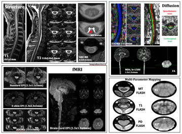
Researcher Kong has been focusing on spinal MRI for a long time. Under 3T field strength, he has optimized a variety of imaging sequences, including 3D T1 and T2 images, gre-me multiple echo structure images, fMRI and DTI with multiple excitation modes, and multi parameter quantitative imaging. Our research group is also committed to optimizing and standardizing the processing flow of spinal cord imaging, especially spinal cord fMRI, correcting EPI image distortion, physiological noise removal, spinal cord image segmentation, individual activation modeling analysis, resting state function connection and network extraction, stable registration and group analysis, finding the most stable analysis method in each processing step will also be a big challenge. The research on neuroscience and clinical diseases in China is mainly focused on brain function, and there is no relevant research on the neuroimaging of spinal cord. The research, development and clinical application of spinal MRI are of great scientific and clinical significance, and have great application potential.
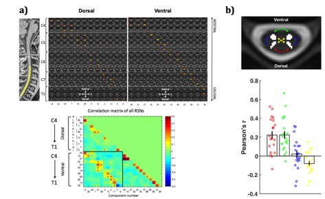
Using the ICA analysis method, researcher Kong found for the first time that there is also a resting network in the spinal cord that conforms to the distribution of physiological characteristics (Fig. a). These resting networks are arranged in the direction of head and tail, each of which is almost no longer than one spinal layer in length. All the resting networks were distributed in the dorsal and ventral gray horn according to the structure of the spinal cord. This functional separation well reflects the neuroanatomical structure of the spinal cord, with sensory processing at the dorsal end and motor signal processing at the ventral end. The ventral network is mainly bilaterally, while the back network is unilaterally. Unilateral dorsal network can reflect hemi body processing, which is consistent with sensory input side (Kong et al. 2014 PNAs). Recently, we also used the seed point functional connection method to verify the physiological mode of fMRI functional connection in resting state of spinal cord under 3T field strength, that is, the correlation between the two ventral horns of spinal cord and between the two dorsal horns, but there was no significant cross functional connection between the dorsal horn and the ventral horn (Figure b) (eippert & Kong et al. 2017 neuroimage).
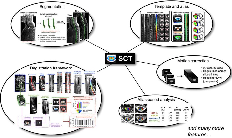
It should be noted that most of the current commonly used brain image analysis software can not be directly applied to the processing of spinal cord image data, because the image characteristics and structure system of the two are often different. SCT is a software package specially used for processing multimodal spinal cord MRI images. It can realize the data standardization of spinal cord images and automatic segmentation, registration, motion correction and other processing steps in the way of code operation. SCT also integrates the latest spinal cord template and atlas, develops the algorithm of spinal cord segmentation and registration based on the template and a variety of analysis methods, which can be obtained automatically In addition, it also includes the preprocessing algorithm for DTI and fMRI of spinal cord. The subsequent steps of physiological noise removal and statistical analysis can rely on FSL and other software. In general, it is a very useful software for the analysis of spinal MRI data.